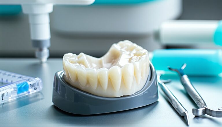
At Reimels Dentistry, we believe that your dental care experience should be reassuring, precise, and empowering. When you or a loved one explores advanced procedures, it is natural to have questions about comfort, accuracy, and safety. One key aspect that can make your visits more transparent is 3d cone beam imaging. This innovative technology provides a comprehensive, three-dimensional view of your teeth, jaw, and surrounding structures. By seeing all the details in a single scan, we can tailor every step of your treatment and ensure you receive the support necessary for lasting oral health. With a commitment to careful and empathetic care, we aim to help you feel confident about the decisions you make for your smile.
However, the benefits of 3d cone beam imaging extend far beyond convenience. From helping diagnose complex root issues to planning intricate restorations, this technology helps reduce guesswork and guide more effective treatment decisions. We understand that you may be curious about the radiation dosage or how this tool compares to more traditional approaches. Throughout this discussion, we will walk you through the fundamentals of 3d cone beam imaging, clarify its unique advantages, and show how Reimels Dentistry strives to make your experience both positive and safe. Your comfort matters to us, and we are here to answer your questions openly so that you can embrace advanced imaging technology with peace of mind.
Explore the basics of 3D cone beam imaging
A revolution in dental imaging
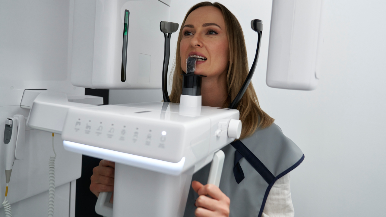
3D cone beam imaging, often referred to as CBCT (cone beam computed tomography), represents a shift in how dental professionals view structures in your mouth. Rather than relying solely on two-dimensional radiographs or typical digital xray imaging, CBCT captures a 360-degree view of the area of interest. According to NCBI Bookshelf, a single rotation of the cone-shaped x-ray beam is sufficient to generate volumetric, three-dimensional images. This difference means that every angle of your teeth, bone, nerves, and soft tissues can be assessed in a single scan, producing detailed information that was once impossible to observe in full.
Scientists and dentistry experts have recognized 3d cone beam imaging as a cost-effective alternative to traditional medical CT scans, mainly because the machines are more compact, the scanning process is shorter, and the overall radiation dosage is lower. These scans are particularly valuable for complex treatments, such as dental implants, orthodontics, and endodontic procedures. By providing a fully realized image of your mouth’s interior, 3D cone beam imaging paves the way for more confident diagnoses and precise treatment plans.
How CBCT differs from traditional methods
You may be familiar with traditional x-rays that display only a flattened, two-dimensional silhouette of a tooth or jaw structure. While these x-rays remain invaluable for routine checkups and simpler diagnoses, they have some inherent limitations. Standard x-rays can obscure smaller or hidden structural anomalies, such as elusive fractures, hidden canals, or problematic bone shapes. CBCT, on the other hand, allows your dentist to study horizontal, vertical, and axial views of every structure in your mouth in a single scan. You end up with a virtual “dental map” that highlights any irregularities in greater detail.
The difference primarily lies in how the image is captured. Traditional medical CT scanners take multiple slices in quick succession, producing immensely detailed cross-sectional images but at the cost of higher radiation. CBCT equipment rotates around your head in mere seconds, gathering numerous images from varying angles. That data is then reconstructed with specialized software, creating a comprehensive 3D model. The streamlined process makes your visit more comfortable, saves time, and potentially minimizes repeated exposures. Moreover, the swift scanning time (often under a minute) helps reduce the risk of distorted images due to minor patient movements.
Consider the benefits for your oral health
Reduced guesswork and enhanced precision
One of the most significant benefits of 3d cone beam imaging is its ability to remove much of the guesswork in diagnosis and treatment planning. Principled decision-making is crucial in dentistry, and having a three-dimensional perspective changes how quickly and accurately concerns can be identified. For example, if you are experiencing persistent toothache and need root canal therapy, CBCT can reveal the exact shape and orientation of root canals, including any narrow or branching canals that might otherwise be overlooked. Studies reported by PubMed Central highlight the superior diagnostic clarity of CBCT in identifying root fractures and resorption, which can directly influence treatment success rates.
This detail-oriented approach also leads to reliable treatment plans. Whether you are considering a cosmetic smile makeover or exploring restorative options, you can rest assured that the recommended procedures are backed by precise and comprehensive information. The 3D images guide your dentist in selecting the right materials, confirming precise angles and depths, and highlighting potential obstacles. This thoroughness reduces the risk of complications during treatment and enhances the likelihood of a positive, long-lasting outcome.
Faster, more comfortable procedures
CBCT not only improves outcomes but also streamlines the care process. Traditional diagnostic approaches might require several rounds of imaging to capture incremental data about different parts of your mouth. By contrast, a single 3D cone beam scan typically captures all the information needed. According to Suwanee Family Dentistry, CBCT scans often produce 150 to 200 images in less than a minute, yielding a holistic view of bone structure, soft tissues, blood vessels, and nerve pathways. Such efficiency can help minimize repeated appointments, saving you time and sparing you unnecessary stress.
Furthermore, the improved accuracy during procedures can help shorten the treatment timeline and reduce your recovery duration. For instance, if CBCT reveals a precise map of your jawbone, the entire process of bone grafting ridge preservation for an implant becomes easier to coordinate. When your dentist knows exactly where and how much bone is needed, the procedure can sometimes be quicker, less intrusive, and deliver more predictable results, helping you feel like your concerns have been thoroughly addressed in the most efficient way possible.
Address concerns about radiation
Understanding the dose
It is perfectly understandable to wonder about the amount of radiation exposure involved in 3d cone beam imaging. The good news is that CBCT generally uses significantly less radiation than a typical medical CT scan. This reduction can be as high as 98% in certain scenarios when compared to older CT technology, according to a study featured by NCBI. That said, the precise radiation dose can vary from 10 to 1000 μSv, depending on the field of view (FOV) and the settings used by your dentist. By carefully selecting the appropriate FOV size—an approach grounded in the “as low as reasonably achievable (ALARA)” principle—your dentist can ensure you receive the smallest amount of radiation necessary for diagnosing or planning your case accurately.
For perspective, the range of CBCT exposure often equates to a few days of background environmental radiation or significantly less than the dose you might receive during a typical hospital-based CT scan. If you have had conventional panoramic x-rays in the past, you can expect CBCT to deliver a somewhat higher dose, but still at a fraction of the exposure used by medical CT imaging of the same region. This balance between clarity and safety is a cornerstone of why many dental practices rely on 3D cone beam imaging for procedures including implants, orthodontics, and TMJ assessments.
Pediatric precautions and ALARA principle
Concerns about radiation become even more important if your child’s dental health requires advanced imaging. Children typically have a higher risk of radiation-induced effects due to their developing tissues and the longer lifetime ahead for potential side effects to manifest. Indeed, PMC notes that children and female patients show a higher propensity for tissue sensitivity, increasing the importance of minimizing exposure.
At Reimels Dentistry, we adhere strictly to the ALARA principle, customizing guidelines for different age groups. CBCT usage is only recommended if the benefit to diagnosis and treatment planning clearly outweighs the risk of exposure. Equipment calibrations can adjust scanning times and lower radiation outputs specifically for children. Our supportive environment also ensures we guide you and your child through the process with empathy and thorough explanations, so that everyone feels safe and well-informed during the imaging.
Use 3D imaging for improved treatment outcomes
Implant planning
Dental implants are one of the most advanced solutions for tooth replacement. They require precise measurements of the jawbone and a complete understanding of nearby structures, such as nerves and sinus cavities. Traditional two-dimensional imaging can measure height and width of the jawbone fairly well, but may miss subtle contours or hidden bone volume issues. A comprehensive CBCT scan, however, accounts for intricate details that might otherwise go unnoticed. By detecting bone thickness, density, and shape, your dental professional can place implants at the ideal angle and depth, minimizing trauma to your mouth and speeding up recovery.
3D cone beam imaging has redefined how dentists tailor implant solutions for each individual. When your practitioner has an accurate 3D map, they can plan for the abutment design and the type of implant that best fits your anatomical profile. The final result is more likely to look, feel, and function like a natural tooth. Additionally, the chance of encountering unexpected complications, such as nerve impingement or poor implant stability, is drastically reduced. According to NCBI PMC, CBCT is now considered a gold standard in evaluating implant sites, precisely because of its capacity for isotropic voxels that generate high-resolution images.
Orthodontic enhancements
If you are thinking about tooth realignment, CBCT can be invaluable for assessing complex orthodontic concerns. Unlike conventional x-rays that flatten the structures, 3D cone beam imaging shows precise root positions, bone levels, and impacted teeth from every angle, helping diagnose associations between teeth and jaw growth. This helps your orthodontist project anticipated movements more accurately, informing decisions such as whether invisalign service might be a suitable option or if other strategies are preferred.
By visually exploring craniofacial structures, your dental team can identify potential issues with the TMJ (temporomandibular joint) that might influence an orthodontic plan. If you or a loved one struggles with jaw clicking, popping, or discomfort, 3D scans can reveal any underlying stress on joints or misguided tooth positions. This synergy between orthodontic realignment and a proper bite alignment fosters more stable, comfortable, and long-lasting post-treatment results.
Endodontic insights
Endodontic work, particularly when dealing with intricate internal canals of teeth, benefits significantly from 3D imaging. Conventional x-rays can show only an approximate outline of canals, which may include superimpositions when multiple roots or teeth overlap. This limited view could mean missing a hidden canal or an early sign of infection. By contrast, CBCT highlights important structural intricacies, helping detect any cracks, anomalies, or branching canals.
If you are scheduled for root canal therapy, you can rest assured that Reimels Dentistry will use every tool available to ensure an accurate diagnosis. A high-precision 3D scan can pinpoint infections such as periapical lesions or inflammatory resorption that might not be visible on a two-dimensional x-ray. By viewing a full, three-dimensional model of your tooth’s interior and surrounding bone, your dentist can shape and disinfect root canals thoroughly, significantly decreasing the risk of reinfection. This attention to detail helps preserve your natural teeth for as long as possible, improving your overall oral health.
Experience personalized care at Reimels Dentistry
Our commitment to supportive, empathetic care
We understand that advanced technology can feel intimidating, especially if you have reservations about additional scans or uncertain results. Our team at Reimels Dentistry is here to offer a supportive environment and clear guidance. Just as different people might need specialized assistance during wisdom tooth removal or tooth extraction, we believe your imaging needs should also be tailored to your specific situation. We know that you may be dealing with emotional stress, and we strive to provide the comfort and attention that make a difference in your experience.
This principle applies to every treatment we offer, from everyday cleanings to comprehensive restorations. If you ever feel apprehensive, our sedation dentistry service can help alleviate anxiety and allow you to stay relaxed during procedures. Our specialized team consistently looks for new ways to reduce discomfort, whether through innovative technology, the latest materials, or a few thoughtful touches in our office environment. We keep an empathetic tone throughout the process, ensuring you feel safe, understood, and empowered.
Combining advanced technology with patient comfort
At Reimels Dentistry, we integrate 3d cone beam imaging seamlessly into your overall treatment plan, balancing precision and safety. Before recommending a scan, we evaluate whether the insights from CBCT would strongly contribute to your diagnosis or treatment. If the answer is yes, we then select the smallest FOV that can still capture the necessary details, thereby minimizing exposure. This rigorous approach makes our practice a trusted destination for individuals and families seeking advanced restorative or cosmetic dental work.
Thanks to CBCT’s swift scanning time and precise results, many procedures—from routine checkups to specialized interventions—are more efficient. If you require a complex restoration or a tooth replacement, advanced imaging creates a clear blueprint for your dentist to follow. For those seeking a full smile rejuvenation, an accurate depiction of bone, gum tissue, and tooth anatomy ensures your cosmetic smile makeover aligns with functional strength and stability. Ultimately, our goal is to help you reach your ideal smile so you feel more confident and comfortable in the long run.
Below is a quick comparison to illustrate how CBCT stands apart from traditional x-rays and medical CT scans:
| Feature | Traditional X-Ray | Medical CT Scan | 3D Cone Beam Imaging |
|---|---|---|---|
| Type of Image | 2D | 3D Cross-sections | 3D Volumetric |
| Radiation Level | Low | Higher | Moderate-Low (10-1000 μSv) |
| Scan Time | Seconds | Several Minutes | 5-40 Seconds |
| Equipment Size | Compact | Large, specialized | Compact, specifically designed |
| Cost | Low | High | Moderate, lower than CT |
| Detail for Dental Structures | Limited | High, but may be excess | High-resolution, targeted at dental |
Such a quick snapshot is just part of the story though, because the real advantage of 3D cone beam imaging is how it facilitates a thorough, holistic look at your oral structures, ensuring no stone is left unturned. For you, that could mean faster appointments, fewer surprises, and the peace of mind that decisions about your dental health are grounded in the clearest available data.
Frequently asked questions
- Does 3d cone beam imaging replace traditional x-rays entirely?
Not necessarily. Traditional x-rays, such as bitewings or panoramic images, remain valuable for routine screenings and simpler diagnostics. However, 3D cone beam imaging is particularly useful when you need a comprehensive view, such as assessing jawbone density for implants or diagnosing complex root canal issues. We at Reimels Dentistry often combine the benefits of both techniques to provide balanced, thorough care. - Will I feel any discomfort during a 3D cone beam scan?
You will likely find the scan process to be quick and comfortable. Most patients only need to bite down on a small tray or positioning guide while the machine rotates around the head. There is no need for bulky sensors inside the mouth, reducing gag reflex worries. The entire procedure often lasts less than a minute. - Is the radiation danger greater for children?
Children are more sensitive to radiation than adults because their tissues are still developing, and they have more years ahead for potential effects to manifest. For that reason, we strictly follow the ALARA principle and tailor the smallest field of view possible. We take extra precautions to ensure that if a child needs a CBCT scan, the benefits strongly outweigh the risks. - How does 3D imaging improve implant placement?
A 3D cone beam scan reveals the precise thickness and density of your jawbone, as well as the location of nerves and sinuses. This knowledge lets us plan the size, angle, and position of a dental implant with greater accuracy, minimizing complications. Ultimately, it helps shorten procedure times, reduce healing complications, and produce a more stable, natural-feeling implant. - Can 3D cone beam imaging assist with cosmetic procedures?
Absolutely. When pursuing cosmetic enhancements like veneers or a cosmetic smile makeover, 3D imaging provides a complete blueprint of your oral structures. This allows for precise, well-coordinated changes that align functionality and aesthetics, helping you get a beautiful result tailored specifically to your anatomy and goals.
By combining the best of modern technology with a supportive philosophy, Reimels Dentistry strives to ensure your procedures are as comfortable and effective as possible. 3d cone beam imaging offers a holistic way to view and plan your dental needs, helping us address unique challenges with greater clarity. From identifying precise root canal pathways to placing implants gracefully, this tool has proven invaluable in crafting personalized treatment strategies.
Our priority is always your safety and satisfaction. This is why every step of our process, from discussing potential benefits to carefully justifying any extra radiation exposure, is organized around delivering comprehensive care in a compassionate environment. We welcome any additional questions you may have about 3D imaging, the breadth of services we provide, or the steps you can take to safeguard your oral health. When you are ready to explore advanced preventive, restorative, or cosmetic dental options, our team is here with open arms, advanced expertise, and the supportive environment you and your loved ones deserve.
